
Anatomy and Physiology
In order to insert, remove and manage chest drains safely and competently practitioners need an understanding of the anatomy and physiology of the chest wall and pleural cavities. A Study has shown that while nurses tend to have a good knowledge of the management of chest drains, they are less clear about the underpinning theory, including the indications for chest drain insertion.
The anatomy of the thoracic wall is important to think about when preparing for chest drain insertion. Between each of the ribs lie the intercostal spaces, which are breached over by the intercostal muscles. Each intercostal space has a nerve, artery and vein running through it. These vessels lie and run just under the rib. This means that a chest drain should be inserted just above the upper border of the rib to avoid damage to the neuro-vascular bundle. The long thoracic nerve runs down the lateral border of the thorax and so chest drains should be inserted anterior to the mid-axillary line to avoid damage to this structure.
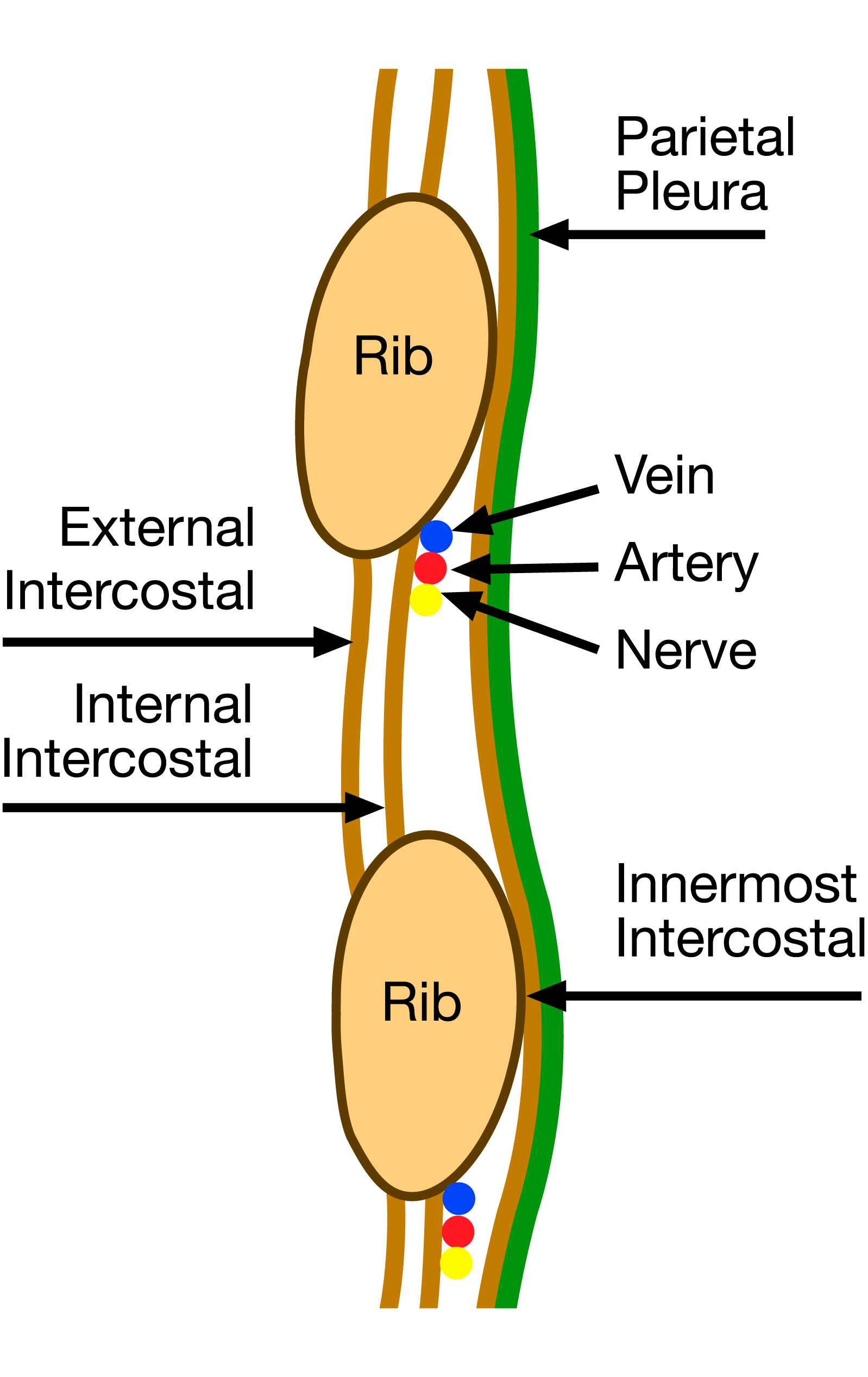
Figure 4. The neuro-vascular bundle inferior to the rib
Remember that the normal lungs are covered with a thin layer of pleural fluid that acts a bit like two wet pieces of glass i.e. they can move sideways but it is difficult to separate them. The intra-pleural pressure in this fluid is below atmospheric pressure, thereby stopping the lungs collapsing away from the thoracic cavity. If air enters the pleural space (pneumothorax) then the lungs will collapse.
For a chest drain to work it must be connected to an airtight seal with an underwater collecting chamber (1,2 or 3 chambers) or a one way (Heimlich) valve with collecting bag. Underwater seal drainage systems are not used in pre-hospital situations. In a patient with a chest drain in place, on expiration, positive intra-pleural pressure is generated. This causes fluid or air in the pleural cavity to be expelled which with an underwater unit will then drain or bubbles one-way through the water. Air passes to the outside air and fluid or blood collects in the bottle. The drain tubing should always be 3 cm below the water level (often pre-marked on bottles). Gravity and differences in pleural pressures between the thoracic cavity and the atmosphere allow this to happen although gravity will work in reverse if the drainage system is lifted above the chest drain when fluid or air will be sucked back into the chest.
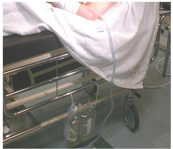 [D]
[D]Figure 5. Showing correct position of drain below patient
1 |
Why should a chest drain always be inserted above rather than below a rib? |